
Feature >
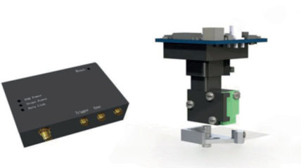
◆System components include microscope body, fixing plate, GRIN lens, CMOS, image acquisition card and acquisition software, steering device, etc.: record calcium signals of a group of neurons at the single-cell resolution level;
◆Suitable for in vivo experiments of freely moving animals;
◆Deep brain imaging can be achieved by implanting GRIN lens;
◆The system is small in size and light in weight and does not affect the free movement and behavioral experiments of mice.
Principle >
◆Preliminary injection of the virus to express GCaMP6 or other calcium ion fluorescent indicators, implantation of GRIN lens and waiting for virus expression
◆Neuron cell activity leads to an increase in intracellular calcium ion concentration, thereby increasing the fluorescence intensity of fluorescent probes such as GCaMP6. The fluorescence is collected by the implanted lens and converted into image signals by CMOS and collected by a high-speed image acquisition card.
◆Image processing software further analyzes the correlation between neuron activity and behavior.


Reviews
There are no reviews yet.