High-speed, high-resolution near-infrared OCT system for animal

High-speed, high-resolution near-infrared OCT system for animal

Optical coherence tomography (OCT) is a high-resolution, non-invasive imaging technology that can produce “super-resolution” images of the eye—without surgery or radiation.  In just a few seconds, it can create high-definition “cross-sectional images” of eye structures, even finer than the thickness of a human hair. This technology is particularly important in ophthalmology, allowing doctors to visualize every layer of the retina, detect early signs of macular degeneration, glaucoma, and other diseases, and even identify eye problems related to diabetes. However, when scientists study eye diseases using small animals like mice, they find that the structure of their eyes differs from that of human eyes.  The eye axis length, pupil size, and other parameters vary, making it difficult for standard OCT devices to produce clear images of the animal retina.

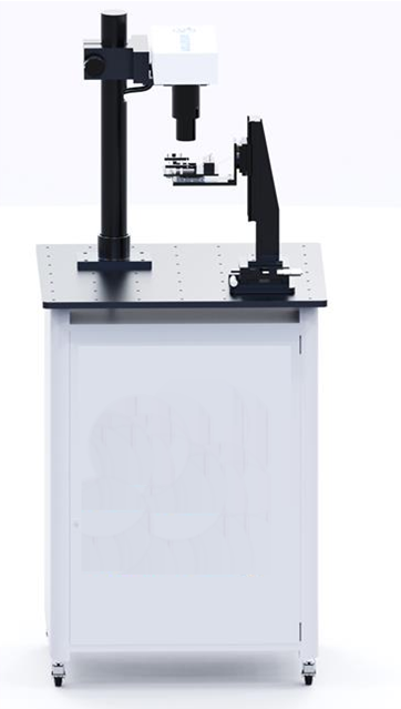
Laboratory near-infrared animal OCT system

  • The 250K scan speed significantly reduces the impact of physiological movements such as respiration and heartbeat in mice, enabling stable acquisition of large-field-of-view OCTA images of the mouse retina.
  • OCTA images allow measurement of various parameters, including blood vessel diameter, vessel density, vessel length density, average vessel tortuosity, and fractal dimension of the vascular network.
  • With an axial resolution of 2.3μm, the system achieves clear imaging of retinal layers, enabling measurement of spectral reflectance between retinal layers and retinal layer thickness.

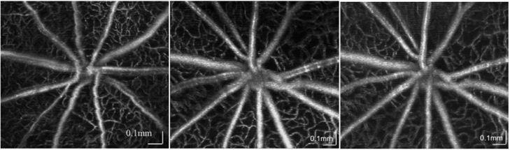
OCTA angiography images
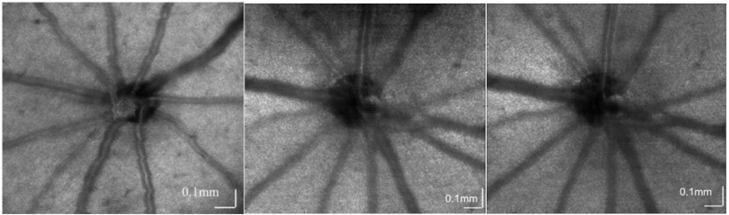
OCT en-face image

Laboratory dual-wavelength animal VNOCT system

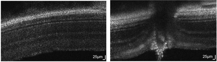
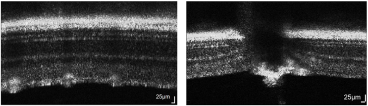
Mouse retina B-scan image


Reviews

There are no reviews yet.

Be the first to review “High-speed, high-resolution near-infrared OCT system for animal”

Your email address will not be published. Required fields are marked *

Shopping Cart
en_USEnglish