


Core Product Advantages
- Olympus Optical Path & High-Fidelity Light Source
Adopts the excellent Olympus optical path to ensure superior light transmittance and imaging clarity. Equipped with a 20,000+ hours long-life high color fidelity LED light source that maintains consistent color temperature at all brightness levels, realizing true color restoration of samples and stable fluorescence excitation for accurate morphological and fluorescence analysis.
- Global Preview for Efficient Observation
Built-in global preview function enables quick browsing of the entire sample slide, helping researchers rapidly locate target observation areas, avoid blind scanning and greatly improve the efficiency of sample observation and scanning.
- One-Click Intelligent Operation
Configured with a high-precision motion platform and electric fluorescence turntable, combined with HDScanner professional scanning software to achieve one-click full-process operation. It simplifies complex fluorescence scanning steps, reduces manual operation errors and lowers the professional threshold for operation.
- Professional Multi-Layer Z-Stack Scanning
Supports advanced Z-stack scanning, seamless image stitching and multi-layer fusion technology, which can capture the three-dimensional structural information of samples and generate ultra-high-definition integrated images. It also supports single-layer image browsing, meeting the needs of in-depth observation of sample multi-layer structure and detailed analysis of local micro-features.
Comprehensive ODM Customization Services
As a professional ODM partner in biomedicine and precision optics fields, HEIDSTAR provides end-to-end customized solutions for this fluorescence imaging system, covering component-level design, mechanical structure optimization and automation control integration. We independently develop high-resolution fluorescence imaging systems and AI-driven pathological scanning platforms for global customers, and offer full-cycle technical support from prototype validation to mass production. We create differentiated microscopic imaging solutions to empower brand innovation and accelerate the transformation from technological concepts to clinical and research implementation.
Diverse Application Scenarios
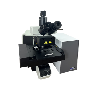
- High-performance Automatic Scanning System
Based on a professional microscope rack design, it is a smart fluorescence microscope suitable for daily work and scientific research in biomedicine, veterinary medicine, microbiology, botany and forensic medicine, meeting the basic and advanced imaging needs of multi-disciplinary research.
- High-throughput Pathological Scanning for Diagnosis
Reliably and repeatedly creates high-quality digital fluorescence slides, with high-speed digital imaging performance and excellent image quality beyond conventional scanning systems. It supports a variety of imaging modes and is suitable for high-throughput pathological diagnosis and fluorescence pathological analysis in clinical departments.
- High-content Screening for Scientific Research
Widely used in cutting-edge life science research fields such as auxiliary drug discovery, cytotoxicity detection, cell tracking, tissue regeneration, cancer research and cell colocalization analysis, providing powerful fluorescence imaging support for in-depth scientific research.
- Morphological & Fluorescence Teaching
With its intuitive operation and excellent imaging effect, it is an ideal teaching equipment for medical and biological majors, helping students quickly master morphological observation and fluorescence imaging analysis skills.
| Parameters | Specifications |
| Loading Capacity | 4 slides |
| Slides Dimensions | 25mm×75mm / 50mm×75mm / 110mm×75mm large slides |
| Bright Field Scanning Speed | 45s (15mm×15mm, 20X objective) |
| Scanning Camera | 5 megapixels Color CMOS |
| Preview Camera | 2 megapixels |
| Objective Lens | Motorized, 20X/0.75NA (4X/10X/40X optional) |
| Pixel Resolution | 0.28μm/pixel (20X) |
| Focus Method | Manual / Auto Focus |
| Barcode Recognition | Supported |
| Multi-layer Scanning | Supported |
| Stage | Motorized XY Stage |
| Image Format | JPEG/HDS/TIFF/BMP/SVS |
| Bright Field Light Source | 3W / 10W LED |
| Fluorescence Light Source | Metal halide lamp |
| Scanning Software | HDScanner multifunctional scanning and recognition software |
Reviews
There are no reviews yet.